Drag the Labels to the Appropriate Locations in This Diagram.
Side with lower concentration of square molecules b. Not all labels will be used Reset Help heps HIV me with a target culpa membrane DNA gones that direct the production of HIV pirtin RNA that direct production of more VISAS 11 recognize proteine on the outside of targut be rds and protect the HIV Dome produces a DNA COPY of the URNA gen Pearson.

Click And Drag Each Label To The Appropriate Dock Chegg Com Plasma Membrane Membrane Structure Cell Membrane
The diagram below shows a bacterial replication fork and its principal proteins.

. Drag the labels to their appropriate locations on the diagram. Use only the blue labels for the blue targets and only the pink labels for the pink targets. Changes in cytosolic Ca 2 concentration link action potentials in the muscle cell to contraction of the sarcomere.
Drag the pink labels to the pink targets to identify processes. Side with higher concentration of square molecules. Drag the labels to their appropriate locations on the diagram below indicating the function of each structure during muscle contraction.
Some atoms hold electrons more tightly than others. Correct Help Reset Help Reset Smooth endoplasmic reticulum ER Central vacuole Nucleus Cell wall Mitochondrion Rough endoplasmic reticulum ER Chloroplast Golgi apparatus Cytosol Cytoskeleton Golgi Apparatus Mitochondrion Nucleus Plasma Membrane. Drag the labels to their appropriate locations in the diagram to describe the name or function of each structure.
5312021 Chapter 16 Lab 3751ANSWERCorrect BioFlix Activity. Glucose released into blood. University of Manchester.
First drag the blue labels to the blue targets to identify the reservoirs in the carbon cycle. Side with higher concentration of square molecules. Drag the blue labels to the blue targets to identify structures.
Drag the labels to the appropriate locations on the diagram of the thylakoid membrane. Identify the structures and determine which hypha is septate and which is coenocytic. Drag the labels to their appropriate locations in the flowchart below indicating the sequence of events in the production of fragment B.
Drag each label to the appropriate location on the. Drag the labels to the appropriate locations on this diagram. Some large molecules move into or out of cells by exocytosis or endocytosis.
Draw the products that would be obtained by following the incorrect arrows in the box entitled A Few Words. Use pink labels for the pink targets and blue labels. Some large molecules move into or out of cells by exocytosis or endocytosis.
Drag the labels to their appropriate locations on the diagram below. Heat sample to separate DNA strands DNA polymerase duplicates DNA strands Cool sample to allow DNA double helices to form Start with a sample of double-stranded DNA. Homeostasis -- Regulating Blood Sugar Both high blood.
One blue target and one pink target should be left empty. Note that although this diagram shows the two types of hyphae a fungus can have either one type or the other but not both Drag the labels to their appropriate locations on the diagram of the fungus and hyphae below. Reset Help actin tropomyosin troponin blocked myosin-binding sites empty Ca 2 binding sites Hint 2.
Drag the labels to their appropriate locations on the diagram of the carbon cycle. Then drag the pink labels to the pink targets to identify the processes in the carbon cycle. HelpReset Low blood glucose Cells in the pancreas Glucagon Liver cells Glycogen breakdown.
How is the sarcomere affected by high or low cytosolic levels of Ca 2. Electronegativity and polar covalent bonds In covalent bonds the electrons are not always shared equally between the atoms. One target of Group 1 and one target of Group 2 should be left empty.
Submit Hints My Answers Give Up Review Part. Labels can be used once more than once or not at all. Filament that pulls the other filament toward the center of the sarcomere.
Note that pol I stands for DNA polymerase I and pol III stands for DNA polymerase III step 2 after pol III binds to. Drag each label to the appropriate location on the diagram. Energy input from the cell.
Drag the labels to their appropriate locations in the diagram. View Available Hint s Reset Help H pumped H diffuses across across membrane membrane site of ATP synthesis site of H release stroma. Drag the labels to the appropriate locations on the diagram of the thylakoid membrane.
Drag the labels to their appropriate locations on the diagram of the water molecules below. Drag the labels to their appropriate locations on the diagram. Energy input from the cell d.
Filament that is pulled toward the center of the sarcomere b. Side with lower concentration of square molecules. Drag the labels to the appropriate locations on this diagram.
One target of Group 1 and one targe. Part A - Animal cell structure Drag the labels onto the diagram to identify the structures of an animal cell.

Layers Of Dermis Layers Of Dermis Dermis Anatomy
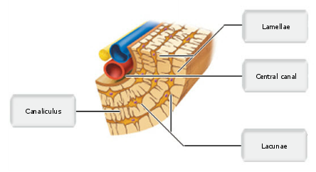
Osteon Labeled Anatomy And Physiology Physiology Anatomy
Skeletal Muscle Physiology Physiology Skeletal Muscle Pearson Education

Print A P Ch23 Digestive System Flashcards Brain Anatomy And Function Liver Anatomy Note Cards
No comments for "Drag the Labels to the Appropriate Locations in This Diagram."
Post a Comment